Blog written by clinical neuropsychologists pertaining to neuropsychological and mental health conditions, diagnosis, treatment and research in Australia.
Monday, 31 March 2014
Life lessons learned from Rake
As I am a newcomer, I'm not completely across the full plotline, characters, subtleties etc., so please forgive any errors. It's not really about the show so much anyway, as about what it can teach us. There's also one correction that 'needs' to be made (we are neuropsychologists after all), to the storyline involving the witness who had sustained a brain injury after a fight with Cleaver Greene.
1. The only certainty in life is change
When I started watching the show, Cleaver Greene was recently out of gaol, had no job, no clients, and no relationship; his apartment had been taken over by his secretary, and his ex-wife was about to remarry. By the end of the series, he had rebuilt his career, retained numerous high profile clients, his ex-wife was single again, and was building what appeared to be quite a healthy relationship with a new woman. Rake takes it to extremes of course, few people's life circumstances are that dire, or change that quickly, but no matter how bad things are, take comfort in the fact that they will change.
2. The brain is an amazing organ
The storyline about Rake's ex-wife Wendy being only able to speak Bahasa following a siege where she is held hostage by her fiancé seems a bit far-fetched, but there have been other cases of individuals losing their first language (temporarily) and only being able to speak a second language following trauma. (As far as I'm aware the other known cases have been following brain trauma rather than psychological trauma, so a bit of poetic licence has been taken by the show here. Or maybe she did sustain a brain injury, I'm not sure.). It is thought that this can happen following damage sustained in the area of the brain where language knowledge is stored. The ability to speak a first language tends to be localised to an area in the left hemisphere of the brain, whereas the ability to speak a second language predominantly comes from the right hemisphere. So when trauma results in better recall of the second language, it may be that they had an injury to the left side of the brain and are better able to access the knowledge of the second language localised to the right hemisphere.
The correction...
The other reference to the human brain in this episode warrants further discussion. Cleaver manages to discredit a witness who had recently sustained a mild traumatic brain injury (mTBI) by claiming that since he had developed amnesia, they couldn't rely on his testimony about events that had occurred (as far as I'm aware) some time prior to the injury. The type of amnesia that follows a mTBI tends to be difficulty recalling events that occurred around the time of the incident. People may experience retrograde amnesia (loss of memories formed shortly before the injury, typically minutes before for a mTBI) and/or anterograde amnesia (difficulties creating new memories after the injury, e.g., remembering new appointments, learning new tasks) following a mTBI. So while it is possible that the witness would not recall that it was Cleaver who attacked him, he is however very unlikely to have difficulties recalling details of the fraudulent deal which was the subject of the court case.
3. Being genuine is endearing
Cleaver Greene is a bit of a 'Warnie' (Shane Warne) type character. No matter how badly he behaves, he always has his fair share of loyal friends and lovers, and the appeal of his character is a big part of the show's success. This is because he is genuine. While he may tell the odd lie to get out of trouble, overall with Cleaver Greene what you see is what you get: he doesn't pretend to be anyone other than who he is. Pretending to be somewhat you're not may impress people in the short-term, but real friendships and relationships are built on a foundation of being genuine.
If you'd like to know more about traumatic brain injury, mental health issues, assessment of brain injury or illness or rehabilitation following brain injury, please contact the team at ANTS.
Friday, 26 July 2013
Protecting your brain's 'borders'
With all the current discussion in Federal politics about 'protecting our borders' it's an opportune time to talk about a less controversial border - the one in our brain. It's called the blood-brain barrier (BBB). Our brain houses up to 650 kilometres of blood vessels! These vessels are lined by a structure, the BBB, which regulates the passage of nutrients, proteins, chemical substances and microscopic organisms between the bloodstream and the brain tissue. Basically, it controls what is, and what isn't, allowed to leave the blood supply and enter the brain.
Blood vessels supply our organs with the nutrients that they need, and help to remove waste products. It has long been known that the blood vessels in the brain and spinal cord are particularly good at protecting the brain from harmful substances, through the BBB, but until recently it hasn't been completely understood how this occurs.
Emerging research using imaging techniques has revealed that the blood vessels in the brain
and spinal cord are lined with specialised cells. These cells have molecular passageways embedded in the cell membrane. These passageways will actively block some chemicals, which are likely to be dangerous to the brain, and play a role in pushing other chemicals across into the brain. The entry and exit of these chemicals are controlled by cells called astrocytes and pericytes.In mouse models, a deficiency in pericytes has been shown to increase the permeability of the BBB to chemicals. And a deficiency in astrocytes has been shown to result in small haemorrhages at the BBB/brain junction.
Using a 'two photon' microscope to probe inside the living brain, researchers have seen these astrocytes and pericytes doing their job, as well as witnessed white blood cells move across from blood vessels into the brain, and back again.
In addition immune system cells, 'microglia', search the brain for harmful substances that have found their way into the brain, and look for damaged or cancerous cells, and remove them.
The practical application of this knowledge is that neuroscientists are now discovering that many neurodegenerative conditions such as Alzhemier's dementia and Parkinson's disease involve defective BBB and related cells.
For example, it is thought that Alzheimer's dementia involves having too much of a chemical called beta-amyloid in the brain. The BBB has a protein that brings the beta-amyloid out of the bloodstream into the brain, and another protein does the opposite. Perhaps keeping the beta amyloid out of the brain might be a prevention, or even a cure, for Alzheimer's dementia.
We look forward to more research in this area, helping us to better understand, and hopefully formulate better treatments for these neurodegenerative diseases.
Thursday, 18 July 2013
See-through brain?
The brain is, understandably, a popular topic for research. Traditionally researchers wishing to study the brain have had to use invasive and time-consuming methods of cutting it up. However, recently a team lead by Dr Karl Deisseroth at Stanford University has developed a method to make brains transparent, allowing them to be studied without having to be sliced up. The process can also be used with other organs, "revolutionising the three-dimensional study of important organs within the body".
The brain is surrounded by lipids that help form the membranes around the cells and bind the tissues together. This membrane blocks the view into the brain. The revolutionary new process referred to as CLARITY replaces the lipids with a clear gel, which preserves the tissues while revealing the internal structure of the brain. The gel is injected into the organ and is absorbed into the tissue. The lipids are then removed, while the rest of the brain remains intact, leaving the neurons, axons, dendrites, proteins and synapses in place and able to be viewed.
Previously these structures could only be seen following the brain being sliced up, and only across tiny slivers of tissue. Researchers then needed to reconstruct three-dimensional data from images of these thin slices to form a three dimensional view of the brain.
Using CLARITY scientists can then use fluorescent proteins and neuro-imaging to see neuronal connections, subcellular structures, proteins, nucleic acids and how cells interact and relate to each other. This opens up the possibility of viewing large networks of neurons with unprecedented ease and accuracy.
The procedure has already been used to investigate the nerves in the post-mortem brain of an autistic boy. They noticed unusual patterns in the neurons, which is likely to help advance the understanding of autism.
We at ANTS look forward to hearing more about how this procedure can be used for advancing our understanding of various neurological and neuropsychological conditions.
Friday, 5 July 2013
Living by values
Values are broad preferences concerning appropriate courses of action or outcomes, which guide ethical action. Commonly held values are honesty, equality, justice and commitment. Value identification, value creation and values-based living are the basis for many religions, philosophies and psychological therapies. A good (though maybe somewhat morbid!) way to identify your personal values is to think about what you would like people to say at your funeral. Or alternatively what you'd like your family to say about you on your 80th birthday, or your partner to say on your golden wedding anniversary.
Values play an important role in good psychological health. In his blog Anger in the Age of Entitlement, Steven Stosny, Ph.D and psychotherapist states that "If you devalue more than you value, your life will be bad, no matter how many good things happen to you. If you value more than you value, your life will be good, regardless of how many bad things happen to you".
That is a very interesting concept, and it explains why:
- Often the things we think will make us happy don't
- Why some people who appear outwardly to have everything are not happy, and
- Why some people who appear to have very little do seem to be happy.
Nelson Mandela is one good example of this. He experienced the death of his father as a child, was expelled from one university and dropped out of another for being a poor student, was imprisoned, experienced the death of two of his children, was not allowed to attend the funeral of his mother and one of his children, and was twice divorced. Despite these setbacks he continued to devote himself to his values of democracy and equality, achieved his goal of democracy in South Africa, and created an important legacy. It is likely that he was able to keep pursuing his goals despite his bad experiences because he was living by his values.
On the other hand, daily we hear about various celebrities, who despite their wealth, status and popularity have drug addictions, convictions for various offences and even on occasion attempt or complete suicide. While we do not know what happens in their personal lives, it is likely that they pursued fame and money thinking that it would make them happy, while ignoring or violating their deepest values. Certainly it is highly unlikely that anyone's deepest values include destroying their bodies and harming other people.
Anything involving the brain that is done repeatedly becomes habituated. Repeatedly pursuing a certain mindset creates neural pathways in the brain that become strengthened until that way of thinking is essentially automatic. If we continue to think about the ways we have been wronged for example, complain about other people and pursue revenge, then resentment will become our automatic way of thinking. If we actively choose to see setbacks as opportunities for growth and pursue compassionate understanding, then we are creating values based thinking, which is likely to lead to more happiness.
The upshot is that values-based living will lead to more happiness and better emotional health. In a practical sense, when something happens in our lives that makes us feel devalued, we are likely to recover more quickly and to have a better overall outcome if we strive to value rather than devalue. For instance, when going through a break up it is better to find ways to feel loved and to love others rather than to blame and demonise the other person.
Certainly easier said than done!
Monday, 22 April 2013
Using neuroimaging to differentiate between Bipolar Disorder and Borderline Personality Disorder
Bipolar Disorder (BD) and Borderline Personality Disorder (BPD) are often confused and misdiagnosed. Both disorders involve dysregulation of emotional responses as key diagnostic criteria. BD is a mental illness involving a set of significant 'mood swings'. The most common form of Bipolar involves 'highs' (or mania) and 'lows' (depression). BPD is a type of personality disorder, or a set of long-standing traits and behaviours associated with significant distress or disability. A key feature of BPD is affective liability, meaning that people with BPD have difficulty stabilising their moods and therefore can demonstrate erratic mood swings. They are also prone to depression. More information about both disorders can be found on the website of the Black Dog Institute.
As it is personality based BDP tends to be a long-term disorder, whereas BD can come and go and with the right treatment can be cured or well managed. Appropriate treatment and management requires proper diagnosis. Unfortunately until now there have been no biological markers for psychiatric disorders.
However, some promising new research has found that there may be a way to differentiate between BD and BPD at a biological level. Professor Gin Malhi from the Sydney University has found biological differences in the brains between people with the two conditions. Professor Malhi and colleagues scanned the brains of people with BD (who were not depressed at the time of the study), BPD and controls with no mental illness while they undertook a task that focuses on emotional responses and is cognitively taxing (the emotional Stroop task). They found a significant difference in the response of the emotional circuits in the brain of the two patient groups and between the patients and controls during the task.
Individuals with BP drew more on the dorsomedial prefrontal cortex of the brain, whilst people with BPD showed heightened activity in the amygdala, a lower level part of the brain which coordinates emotional responses and processes negative emotions such as fear. Professor Malhi reported that people with BPD were not able to regulate this "key node in the lower part of the brain that co-ordinates emotional understanding". In contrast, people with BP were able to function quite normally when well, and just needed to expend more effort in the brain than controls.
While the results are very promising, Prfessor Malhi stresses the need for replication of the study.
Friday, 5 April 2013
Right brain/left brain
If further proof was needed that the human brain is a fascinating organ, because of the way our brains are organised, there is a slight but discernible difference between the left and right sides of our faces.
Our brains are composed of approximately 100 billion neurons (nerve cells), with 50 billion neurons in each half. However, only approximately 250 million neurons join the two halves, meaning that our left and right brains are mostly unconnected and work separately. One result of this is our faces are unsymmetrical.
Many actors and other famous people (such as David Caruso, pictured) are known for having a 'good side' - preferring to be photographed on one side of their face than the other. Research shows support for the assertion that the different sides of our faces are different and one side is actually considered to be more attractive than the other.
Professors Kelsey Blackburn and James Schirillo from the University of Wake Forest asked undergraduate university psychology students to rate the attractiveness of 20 volunteers who had been photographed on both their left and right sides, about 37 degrees from the centre on each side. Some photographs were untouched, while other photos has been Photoshopped so that they were mirror-reversed so the left side looked like the right and vice versa.
As well as asking them about their preferences, the researchers examined the students' pupils. The size of one's pupil is related to the intensity of our emotional response to stimuli. In general, the students preferred the left face of the both men and women — regardless of whether they were shown the untouched left face, or a left face which had been mirror-reversed to look like a right face. This was regardless of the gender of the person in the photo, and the gender of the person doing the judging. And the pupils of the volunteers opened larger when they rated the faces more attractive.
The echoism app, developed by artist Julian Wolkenstein shows a combined image of two photos of either the left or right side of people's faces, with most of these photos posted on line being very different from the people's actual image. This indicates that 'symmetrical' faces don't look quite right. Judge for yourself whether you think the right or left images are more attractive.
Wednesday, 13 March 2013
The psychopathic brain
A very interesting article by Dr Karl Kruszelnicki revealed that psychopathology, characterised by features such as egocentricity, a lack of empathy, remorse and poor impulse control, may be associated with the orbito-frontal cortex in the human brain. The orbito-frontal cortex (OFC) is located in the pre-frontal area of the frontal lobes behind the eyes, as indicated by the green shading on the image to the right.
The OFC controls those behaviours which are known to be impaired in psychopaths, including impulse control, planning and calculating risk. It also has a role in processing odours, and research has shown that psychopaths have a poor sense of smell.
Shamay-Tsoory, Harari, Aharon-Peretz, and Levkovitz (2010) proposed that psychopathy is associated with impairments in theory of mind (TOM) which is also associated with the OFC. TOM is the ability to understand what another person may be thinking and feeling, and to be able to appreciate their perspective. Shamay-Tsoory et al. showed that people with lesions in the OFC performed poorly on complex TOM tasks, particularly those drawing on affective TOM, which involves interpreting emotions. People with psychopathic tendencies had similar difficulties on complex TOM tasks as those individuals with lesions in the OFC.
The authors suggested that psychopaths may not appreciate the emotions, fears and sorrow of other people, which allows them to behave callously without feelings of remorse.
The OFC also has extensive projections to a part of the brain called the amygdala. The amygdala is involved in processing fearful and sad facial expressions and emotions and has a role in instrumental learning. Several neuroimaging studies have shown that people with psychopathic tendencies have reduced amygdala volume (Kiehl et al., 2001; Tiihonen et al., 2000).
References
Kiehl, K.A., Smith, A.M., Hare, R.D., et al. (2001). Limbic abnormalities in affective processing by criminal psychopaths as revealed by functional magnetic resonance imaging. Biological Psychiatry, 50, 677-684.
Shamay-Tsoory, S. G., Harari, H., Aharon-Peretz, J., & Levkovitz, Y. (2010). The role of the orbitofrontal cortex in affective theory of mind in criminal offenders with psychopathic tendencies. Cortex, 46, 668-677.
Tiihonen, J., Hodgins, S., Vaurio, O., et al. (2000). Amygdaloid volume loss in psychopathy. Society for Neuroscience Abstracts, 2017.
Tuesday, 26 February 2013
Sports stars: a great example of brain-behaviour relationships
New research has found that athletes have a superior ability to rapidly process pictures, and to quickly learn unpredictable and dynamic visual information. This ability appears to give them the edge on the sporting field (or court..or arena).
In a study conducted by Jocelyn Faubert and colleagues from the University of Montreal, 102 professional athletes, 173 amateur athletes and 33 non-athletic students were asked to track and describe a series of abstract moving pictures on a screen. This was repeated 15 times over five days. The task was deliberately neutral and unrelated to sport, so that the results could not be assumed to be related to the athlete's greater familiarity with the tasks.
The professional athletes processed the visual information much faster than the other groups and improved their performance markedly over the five days. The other two groups started similarly, but the amateur athletes soon improved and had a much faster learning speed than the non-athletic students.
The results are a great addition to our understanding of the relationship between sporting prowess (behaviour) and cognition (brain), because previous studies have not found a significant relationship between sporting ability and performance on other cognitive tests.
This study revealed that "[Professional athletes] appear to be able to hyper-focus [on complex visual information] for short periods of time resulting in extraordinary learning functions," according to Jocelyn Faubert. The cognitive requirements for interpreting the visual information parallel situations such as driving, crossing the street and perfoming sport activities. Overall, they are smarter at learning how to interpret the "real world in action".
This coincides with previous research which showed differences between athletes and non-athletes in a part of the brain which regulates motion perception.
The study did not explore whether these abilities were innate or acquired through practice. This would be an interesting area for further research.
Monday, 11 February 2013
How the brain organises our visual world
We live in a world in which we are bombarded with thousands of visual images every day. Recently researchers at the University of California provided a unique insight into how the brain organises and stores these visual images.
In the study, subjects watched hours of movies which included scenes from everyday life, whilst their brain activity was captured using functional magnetic resonance imaging (fMRI). They collected imaging data on 1,705 categories of objects (e.g. dog, building, road, furniture) and actions (e.g. jump, spin, hit, touch).
They then created a map to show how images of these objects and actions were organised by the brain. What they found was that the brain works efficiently to categorise images enabling similar images to be stored together in compact brain regions.
The graph below, taken from this study, shows how the brain links categories which are semantically related. To make it easier to identify, researchers have shown similar categories using the same colour. For instance, images to do with “person” are shown in green, whilst animal images can be seen clustered together in yellow. Images related to vehicles are identified by pink.
From this we can see how the brain links related categories together. Categories that are represented similarly in the brain are plotted at nearby positions. For example, information about humans shares the same neighbourhood in the brain as information about animals. Categories that have less in common, however, are located further away from each other in the brain. The graph below shows how to the brain, “person” and “talking” are represented as being more similar and having more in common than “kettle” and “talking”.
©University of Berkley
This study suggests that rather than each category being stored in its own distinct brain region, the brain is able to determine whether or not diverse categories share overlapping or common features and then group them accordingly in a continuous space. The brain actively ascertains the relationships between categories in order to work out where to store images. This more effectively utilises the relatively limited brain space available, given the size of our brains, and helps the brain to be more efficient by minimising the number of neurons required to represent each feature of an image.
A further interesting finding was that whilst only five subjects were included in the study, the authors were able to ascertain that different people share similar semantic layouts. That is, all of the subjects tended to use very similar cortical maps to store visual images. As well as providing us with a unique insight into brain organisation, the results of this study have implications for improving computer image recognition systems and creating other brain-machine interfaces.
Researchers have produced an interactive version of the brain map which provides a detailed insight into the visual function and organisation of the brain which can be found by following the link: http://gallantlab.org/semanticmovies/
Tuesday, 22 January 2013
An update on Molly Meldrum
Molly's recovery
Readers may recall that in December 2011 Molly Meldrum was admitted to hospital after a fall at his home in Melbourne. He was in an induced come for approximately 10 days and in Post Traumatic Amnesia (PTA) for almost one month. From the limited details released by the media, Molly appeared to have sustained at least a severe Traumatic Brain Injury (TBI).
It has emerged that on February 27, 2012, less than 10 days after coming out of PTA, Molly checked himself out of hospital. At that point he could walk and talk, was physically much better, had likely had enough of being in hospital and (incorrectly) assumed that he was well enough to go home.
Friday, 11 January 2013
Should we be concerned about the effects of mobile phones?
Numerous articles and studies have suggested that regular use of mobile telephones may be harmful to our health. It has even been suggested that mobiles fry our brains. Given that mobile telephone are becoming more and more prevalent, are being used by younger and younger children, and for many of us are our primary means of communication, it is important that we understand the effects of these devices on our brain functioning. It is also important that we do not make false assumptions about the erroneous effects of mobile phones.The public concern about mobile phones is based on the fact that wireless phone signals cause electromagnetic fields from which radiation is emitted. The World Health Organisation has found that radiation is a possible human cancer-causing agent.
It is however difficult to measure to what extent radiation from mobile phones heats human brain tissue. A recent study by researchers from the Memorial Sloan-Kettering Cancer Center and the Alcatel-Lucet Bell Laboratories in the US found that MRI scans can directly study how mobile phones heat the human brain. Research is in its early stages however and there are no conclusions as yet.
Wednesday, 14 November 2012
Non-pharmacological Treatments for Memory Decline
It was recently reported on ABC news (Mon Oct 15, 2012) that drug companies are abandoning research into the development of new drugs for dementia.
The key reasons cited for this trend were the enormous costs involved in drug development and little success with disease modifying drugs, particularly for Alzheimer's Disease, the most common form of dementia.
With dementia widely recognised as a ballooning problem for not only in Australia but across the world over, this then raises the question of what other treatment options are out there?
In fact there are several other potentially promising lines of research investigating non-pharmacological treatments for memory decline. These non-drug based treatments share one thing in common, capitalising on the neuroplastic and regenerative properties of the brain.
One such treatment which has gained increasing attention is cognitive remediation, a psychological intervention which has been particularly investigated in people with schizophrenia. This form of intervention typically involves the use of brain training exercises and teaching of memory strategies, with research overall showing robust benefits for memory and cognition, as well as improvements in quality of life. Cognitive remediation has additionally recently been trialled in people at risk for dementia, with early promising results showing improvements in memory.
Another exciting new intervention is through using mild forms of brain stimulation. These brain stimulation techniques involve the application of small electric currents or brief magnetic pulses into the brain for the purpose of transiently modulating brain activity. These non-invasive forms of stimulation have been shown to be effective in treating depression, and there is now some preliminary research showing that they can also be used to enhance memory in older persons and people suffering from Alzheimer's Disease.
Though the research into these new non-pharmacological treatments to help improve memory is very much preliminary at this stage, with more research these treatments may become more widely available in the future. Most likely these new treatments will be used to supplement existing drug treatments, as additional “booster” treatments to help delay cognitive decline and improve quality of life.
ANTS conducts a Cognitive Remediation Programme based in Sydney's inner west. For more information please contact us.
Researchers at the University of New South Wales are also currently conducting a research trial investigating the use of mild brain stimulation for people at risk for dementia. More information about this trial is available at http://www.cheba.unsw.edu.au/project/brain-stimulation-and-cognitive-training.
Monday, 5 November 2012
An unconventional treatment for dementia?
Curry may help combat dementia!
Scientists are investigating whether curcumin, a chemical in the spice turmeric, may help to prevent Alzheimer's dementia (AD), the most common form of dementia. Curcumin, a powerful antioxidant and anti-inflammatory, may play a role in clearing the brain of a protein called beta amyloid, which is known to be a causative factor in the development of AD.
India, the largest consumer of turmeric-based curries has the lowest rate of AD in the world.
AD and other forms of dementia are perhaps the single greatest health issue facing Australia in the 21st century, with almost 1 million people in Australia predicted to be living with dementia by 2050. A number of researchers and medical practitioners have recently spoken out about their concerns about the reduction in research dollars spent on finding ways to combat this insidious illness, given the morbidity and prevalence of dementia. Many pharmaceutical companies have stopped or reduced their dementia research programs due to the lack of efficacy of treatments to cure the disease or slow its progress.This is despite advances in neuroimaging technology and diagnostic techniques.
As with most illnesses, prevention is better than cure, and scientists are now focusing on finding ways to stop those at risk of developing AD from doing so.
A clinical trial has started at a Sydney retirement village where 100 residents will take supplements of curcumin and be given MRIs. Ralph Martins, a professor of ageing and Alzheimer's disease at Edith Cowan University, who is conducting the research along with the McCusker Alzheimer's Research Foundation, the Brain and Mind Research Institute and the Royal Prince Alfred Medical Imaging Services, said that:
While the research is promising, there are a range of other lifestyle factors, including diet, exercise and 'brain training' (or stimulation to keep the mind active) that are also important to consider in trying to prevent all forms of dementia.
Curry is certainly not enough, but it may be a good start!
If you are concerned that you or a loved one may be developing dementia, we at ANTS can help. The first step is to read our fact sheet on dementia at:
http://neurotreatment.com.au/neuropsychological-information-dementia.aspx
A thorough neuropsychological assessment is a key component of the diagnosis and management of dementia. If you would like to learn more about what we can offer here at ANTS, please don't hesitate to contact us.
Monday, 22 October 2012
The Neuropsychology of Depression
In his recently released autobiography, Ian Thorpe describes how he has suffered from depression most of his life. Like many young Australians with depression, the 30-year old revealed that he kept the illness behind closed doors to the point that not even his family and close friends were aware of his battle with the mental illness, and rather than seeking professional help he would instead often turn to alcohol. He recalls in his book periods of low mood that would often last a month, during which he contemplated suicide.
Depression is not uncommon in young Australians, with approximately 160 000 individuals between the ages of 16 to 24 years of age living with the mental illness. Like Thorpe, a high percentage of sufferers experience suicide ideation and such individuals are at increased risk of completing suicide.
Depression is mainly known for its affect on mood. A lesser known fact is that sufferers also exhibit a number of neuropsychological impairments. Research has shown that individuals with depression have poorer episodic memory and slower psychomotor speed. In addition, those with depression exhibit difficulties with problem-solving, planning and mental flexibility.
The range of cognitive impairments seen in individuals with depression is the consequent of several biological changes in the brain. One of the most widely researched biological dysfunction concerns a deficiency in Serotonin, an important neurotransmitter that is known to mediate memory and learning. Brain MRI studies have shown that, relative to healthy controls, individuals with depression have decreased activity in the frontal lobe, an areas of the brain important for problem-solving, planning and mental flexibility. In addition, research has shown that depressed people exhibit abnormal activity in the amygdala, an area important for processing emotional aspects of information.
Thorpe described that after years of suffering with depression he finally consulted with his doctor who prescribed medication “but little in the way of explanation”. Ironically, he noted that this consultation isolated him further as he felt he "now had a secret and no one to share it with.”
While most antidepressant medications are thought to improve symptoms of depression by correcting deficiencies in Serotonin, they are not affective in addressing the important psychological component of the mental illness, namely negative and maladaptive thinking-patterns. This is usually addressed by supplementing antidepressants with some form of cognitive therapy. Interestingly, there is some preliminary evidence to suggest that cognitive therapies may in themselves alter neural circuitry and normalise abnormal amygdala and frontal activity. Whether these changes lead to corresponding neuropsychological improvements, however, is yet to be determined.
Monday, 27 August 2012
Modern day Phineas Gage
As a psychology student one of the first things you learn about in class is the classic case of Phineas Gage, a 19th century construction worker who was unfortunate enough to have an iron rod driven completely through his head, damaging a substantial part of the left side of his brain, and surviving the whole ordeal.
Phineas Gage, who’s injury resulted in damage to the left frontal lobe, is often documented to have been left with profound behavioural and personality changes, not surprising given that the frontal lobe is responsible for higher mental functions important for social situations, such as reasoning and decision-making.
The case is significant in the field of neuropsychology because it influenced early discussion (and findings) that damage to specific parts of the brain can affect specific functions while leaving other functions intact. The case of Phineas Gage has also been popularised in the media, being mentioned in episodes of shows such as House, Medium and Ripley’s Believe it or Not (Fridge Magnet anyone?).
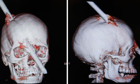
Amazingly, a 24-year old Brazilian construction worker recently sustained a very similar injury (though the injury was to the right side), and like the classic case, the worker was not only conscious moments after sustaining the injury but he was also able to talk and explain to doctors what happened. The worker reportedly felt little pain and looked as if nothing had happened following the accident.
As can be seen from the photo, the iron bar (which had fallen from the fifth floor of a building) entered the back of the worker’s skull and exited between his eyes. He reportedly narrowly escaped losing one eye and permanent left-sided paralysis.
The doctors who treated the Brazilian worker remarked that the iron bar entered a “non-eloquent” part of the brain that does not have a specific known function. Though, with the patient reportedly still in hospital for at least two more weeks, only time will tell as to whether this man will ultimately develop any of the long-standing behavioural and personality changes sustained by his 19th century counterpart.
As argued by Dr Hamberger, Clinical Neuropsychologist at the Neurological Institute of New York, the lack of reported concern by the worker that a 1.8 metre long iron bar has penetrated his head may be evidence in itself of more subtle higher-level cognitive changes that won’t be immediately apparent before more detailed neuropsychological testing, such as self-awareness and insight.
Wednesday, 11 July 2012
The neuropsychology of smell
The power of the nose
Have you ever smelt something that immediately took you back to your childhood, to a happy or difficult time in your life or to a certain place? Could you remember exactly how you felt or what you were thinking or doing when you last smelt it, even if it was a long time ago?
It's a common experience to associate an aroma with a memory, which can often arouse quite vivid feelings. The reason that our sense of smell is so powerful is that unlike other senses it does not have to pass through a brain structure known as the thalamus but is directly processed by the olfactory system in the brain. It is also part of the limbic system, which also plays a role in processing emotions and memories.
Compared to many animals, the human olfactory system takes up a relatively small part of the brain and our sense of smell is quite weak. However, the importance of human olfaction is demonstrated by studies that show we can us it among other things, to identify relatives, choose partners and determine age
A new study by Assistant Professor Johan Lundström, an experimental neuropsychologist at the Monell Chemical Sciences Centre in the United States, demonstrates how well we can use our sense of smell to determine age. And debunks the myth that old people smell bad!
Assistant Prof Lundström and colleagues had people from three age groups (20-30 years, 45-55 years and 75-95 years) sleep with pads located under their arms for 5 nights. The pads were then placed in jars and other participants were asked to group the jars according to age. Participants were able to easily group the armpit pads by age group and even by gender.
Surprisingly, despite the common conception that older people have an unpleasant odour, younger people rated the arm pads of the oldest age group as being fairly neutral and not unpleasant.
The group rated as most unpleasant was men aged 45-55 years.
The work of neuropsychologists is varied and valuable. To learn more about what we do and about our services, visit our website
Friday, 25 May 2012
Research reveals the part of your brain responsible for a song getting stuck in your head!
If you've ever had a song stuck in your head, you'll be interested in the latest research investigating the brain region responsible for recognition of familiar tunes revealed as part of a study into Semantic dementia.
Semantic dementia research
Semantic dementia (SD) is a rare type of dementia characterised by a loss of semantic memory, memory for generalised concept based factual information (i.e, general knowledge). The most common symptoms, particularly early on, are an impaired ability to retrieve words from memory and impaired comprehension of word meanings. It also affects patients' ability to recognise objects and sounds.
Wednesday, 16 May 2012
Parenthood changes your brain!
Parents may not be surprised by the finding that being a parent actually changes your brain (some may be surprised that the changes are good ones!). Research shows that parents have different patterns of brain activity in some areas than non-parents.
One study found that hormonal changes that occur in pregnancy increase the number of dendrites in the brain. Dendrites are the projections from a neuron (or brain cell) that act to facilitate communication between neurons. So, the more dendrites, the more chance of neural connections. The neural connections that are created during pregnancy have been shown in rat studies to enhance memory and learning. This is likely to be because learning and memory are important in raising young.
Thursday, 10 May 2012
Mental health and the federal budget
The 2012-13 federal budget has been released, and according to some expert commentators, there is good and bad news for mental health arising from the budget.
The good news is:
- Overall funding for mental health has not been cut.
- Funding for the Mental Health Nurse Incentive Program, providing incentives for community based general practices to engage mental health nurses, has been maintained.
- The government will provide $21 million to fund additional allied mental health services for patients under the Better Access initiative, whereby treatment for those with diagnosed mental health problems is partially covered by Medicare.
- New spending of $115m over five years is directed towards better mental health care for veterans.
- From 2014-2015 $9.1m will be provided to the Australian Bureau of Statistics to conduct its Survey of Disability every three years, rather than every six years.
Monday, 7 May 2012
Positive thinking changes your brain!
Psychology has for some time now borrowed from the philosophies and practices of Buddhism (e.g., meditation and mindfulness), to help treat conditions like depression and anxiety and to help people increase their general psychological well-being.
New breakthroughs in neuropsychology and neuroscience have allowed us to better understand how these practices work to actually rewire the brain to allow for more calmness and greater happiness.